Home

Joseph Banks Concurso letal cunean tendon horse ensillar Omitir Menos que

Image: Anatomy for tarsus nerve block, horse - Merck Veterinary Manual

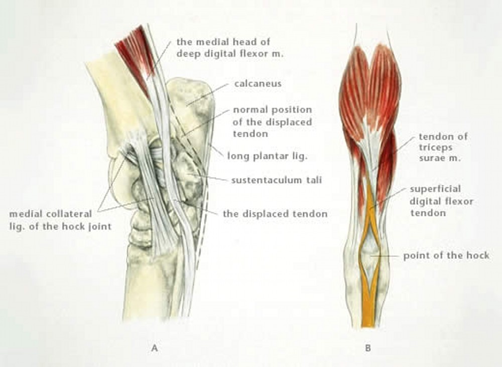
Luxation of the Superficial Digital Flexor Tendon from the Tuber Calcanei  in Horses - Musculoskeletal System - Merck Veterinary Manual
Luxation of the Superficial Digital Flexor Tendon from the Tuber Calcanei in Horses - Musculoskeletal System - Merck Veterinary Manual
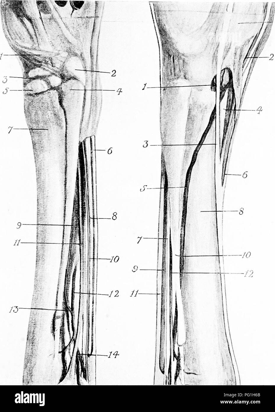
Image: Arthroscopy joint entry sites, hock, horse - Merck Veterinary Manual

IHMC Public Cmaps (3)

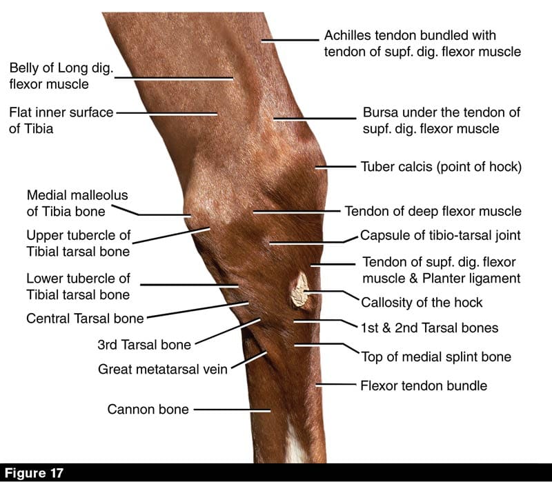
Hock Provides the Horse Thrust Under Immense Strain

Causes of Equine Lameness | EquiMed - Horse Health Matters

Review of Equine Distal Hock Inflammation and Arthritis | Semantic Scholar

GALLERY2: EQUINE STUDIES | Jayhawkartandimage

The Project Gutenberg eBook of Lameness of the Horse, by John Victor Lacroix

Gastrocnemius Tendonitis - ScienceDirect

If Only They Could Talk

2.8 Hock Lameness Flashcards | Quizlet

The surgical anatomy of the horse ... Horses. Plate XXVIII.—Metatarsal  Region, showing Arteries, Tendons, Ligaments, Bones, etc. A.—INNER aspect  I. Cunean tendon. 2. Cuneiform parvum. 3. Scaphoid. 4. Head of inner
The surgical anatomy of the horse ... Horses. Plate XXVIII.—Metatarsal Region, showing Arteries, Tendons, Ligaments, Bones, etc. A.—INNER aspect I. Cunean tendon. 2. Cuneiform parvum. 3. Scaphoid. 4. Head of inner
Ungulate Anatomy Lab 7

Hock Provides the Horse Thrust Under Immense Strain

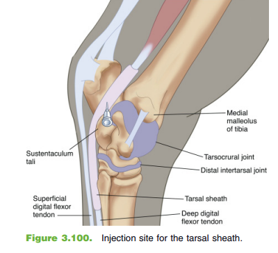
How to Inject the Tarsal Joints

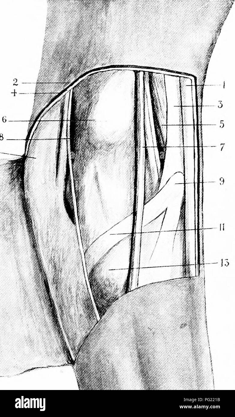
The surgical anatomy of the horse ... Horses. ,.>10- t: 'S. Plate  XXI.—Posterior Tibial Nerve and Cunean Tendon A.—posterior tibial ner'e and cunean  tendon exposed I. Posterior tibial nerve. 2. Tendo-Aciiilles.
The surgical anatomy of the horse ... Horses. ,.>10- t: 'S. Plate XXI.—Posterior Tibial Nerve and Cunean Tendon A.—posterior tibial ner'e and cunean tendon exposed I. Posterior tibial nerve. 2. Tendo-Aciiilles.

Lameness Flashcards | Quizlet

Tendons - Horse Anatomy - WikiVet English

Retinaculum: The term retinaculum... - Fox Run Equine Center | Facebook

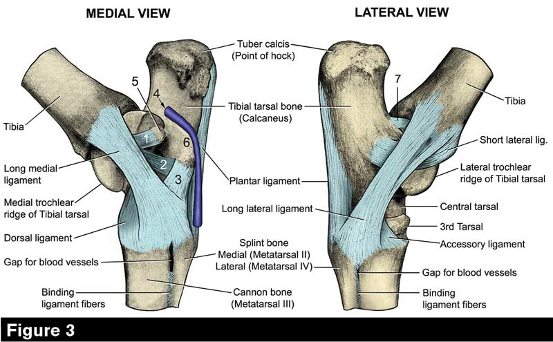
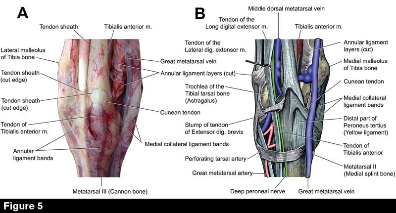
Tarsal Anatomy of the Horse

The topographical anatomy of the limbs of the horse. Horses; Physiology.  126 TOPOGEAPHICAL ANATOMY OF M. adductor. M. sartorius. -  semimeml:iranosus. M. semitendinosus. M. gastrocnemius. A.saphena. -. X.  saphena. i[. popliteiis ]
The topographical anatomy of the limbs of the horse. Horses; Physiology. 126 TOPOGEAPHICAL ANATOMY OF M. adductor. M. sartorius. - semimeml:iranosus. M. semitendinosus. M. gastrocnemius. A.saphena. -. X. saphena. i[. popliteiis ]

Tarsal Anatomy of the Horse

Gastrocnemius Tendonitis - ScienceDirect

How to Inject the Tarsal Joints

Hock Provides the Horse Thrust Under Immense Strain

How to Inject the Tarsal Joints